Welcome to Virtual Embryology!


What is Virtual Embryology


@@@In the Congenital Anomaly Research Center of Kyoto University, approximately 1000 specimens are maintained in serial sections. This fetal library is used to establish normal standards of human preneral development and to find the real incidence of anomalies. The system, Virtual Embryology, is designed to help understanding the structure of embryo using 3D image reconstructed from these microscopic serial sections. A user can "dissect" the virtual embryo on a computer display and browse it from arbitrary viewing angle. Also an animation of virtually growing embryo can be shown in this system.


Why Virtual Embryology?

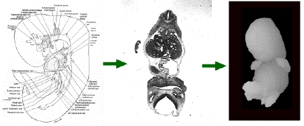
Complicated scheme?? Microscopic section? 3D image!


See how a heart grows inside a embryo. Click here! (1022KB MPEG movie)


How these images come

Equipments used
Reconstruction process of 3D embryo


What you can see here

3D surface view
stage 15: whole body (763KB MPEG movie)
stage 15: heart (690KB MPEG movie)
stage 18: heart (753KB MPEG movie)
stage 20: whole body (746KB MPEG movie)

Resected view

"Interactive resection" has not been impremented on this page yet.
But on a standalone system(Mac + NIH Image), it works as this.

Virtual growth

To see the Growing Virtual Embryo, click here! (161K MPEG movie)


Who made this

Takashi Miura, M.D.
Assistant Professor, Department of Anatomy and Developmental Biology
Congenital Anomaly Center
, Faculty of Medicine, Kyoto University
Re: Whole research (NIH Image's macro programming, Data acquisition, etc)

Masaru Komori, Ph.D.
Professor, Computational Biomedicine, Shiga University of Medical Sciences
Re: Homepage author, Technical management

Kohei Shiota, M.D.
Professor, Department of Anatomy and Developmental Biology
Chief Chair, Congenital Anomaly Center
, Faculty of Medicine, Kyoto University
Re: Anatomical verification


Reference

Miura T, Komori M, Takahashi T, Shiota K. 1995. Computerized Three- Dimensional Reconstruction of Human Embryos and Their Organs Using the "NIH Image" Software. Acta Anatomica Nipponica 70:353-361.

Komori M, Miura T, Shiota K. 1994. Three Dimensional Reconstruction from Human Embryo Sections and Animated Presentation of its Development Process. Medical Imaging Technology 12:493-494.

Komori M, Miura T, Shiota K, Minato K, Takahashi T. 1995. Virtual embryology: a 3D library reconstructed from human embryo sections and animation of development process. Medinfo 2:1229-30.